Reimagining medical diagnostics in developing countries: meta-optics offers a new way to look inside cells
24 Aug, 2021
Developing countries face many challenges in the fight against infectious diseases. One of these is the lack of access to medical diagnostic tools. This is considered a key reason why malaria and tuberculosis are still two of the leading causes of death in sub-Sahara Africa and parts of Asia.
In order to address issue of affordable, accessible medical diagnostic tools, new technologies are needed. That’s where nanotechnology comes to the rescue.
The study of biological cells is essential to medical diagnostics, disease detection and prevention. Traditional pathways to ‘see’ cells, their internal structures and the interaction between different organelles, involve staining the cells with special dyes and using phase contrast imaging. These established techniques deliver a great deal of information, but the colored dye can kill it. The phase contrast microscopes are bulky and expensive instruments that have optical parts that need maintenance over time, making wide-spread use difficult in low income countries.
New research is being done in an effort to address these challenges. Since cells are so tiny (on the order of a few to 100 micrometres or so), it makes sense to create ‘eyes’ to see on that level using nanotechnology.
Currently, it is possible to fabricate devices and nanostructures with unparalleled precision. Application of such nanostructures to ‘play’ with light has led to the emergence of a new field of optics known as meta-optics (meta stands for beyond in Greek) that aims to miniaturize and condense the current bulky microscopes into something more portable and cost-effective, without affecting the performance. To that end, meta-optics can be of utmost value in making medical diagnostics more accessible. If that sounds like sci-fi, it is not.
As a matter of fact, that’s what Professor Ann Roberts from the University of Melbourne and her team have done. Using cutting-edge nanotechnology, they have designed an array of nanostructures on a conventional microscope coverslip that enable the user to see the transparent biological cells, without staining. Not only does this not harm the cells, it also incorporates the phase contrast microscope’s ability to visualize transparent cells without the need for the phase microscope itself— a win on both fronts.
Prof Roberts says, “Our approach has significant potential to become an inexpensive, ultra-compact phase-imaging tool that could be integrated into smartphone camera and other mobile devices to make mobile medical diagnostics broadly available.”
Details of this nanophotonics enabled coverslip (NEC) and its ability to visualize micrometer-sized phase components were recently published in Light Science and Applications.
To demonstrate the working of the device, human cancer cells (HeLa) were placed on the NEC. A beam of light was passed through the cells on the NEC and onto a camera to capture the image. By tilting the NEC by a small degree, the phase contrast of the cells was dramatically enhanced to produce a clear visualization of the cells, which were otherwise not clear.
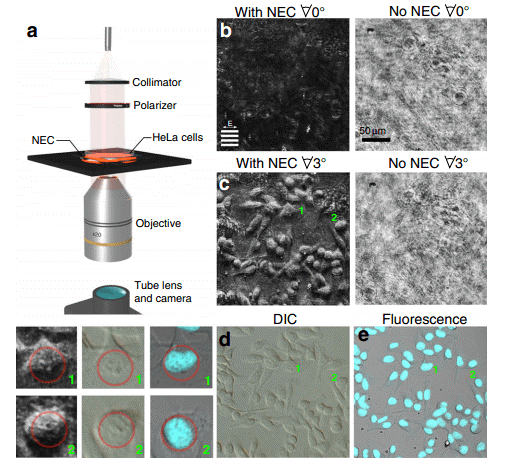
Image credits: Lukas Wesemann, Jon Rickett, Jingchao Song, Jieqiong Lou, Elizabeth Hinde, Timothy J. Davis, and Ann Roberts
Unlike a differential interference contrast (DIC) microscope that uses bulky Wollaston prisms for imaging, the coverslip is much smaller, yet creates a similar three-dimensional contrast effect. The NEC achieves this using the synergy of an optically active layer and fine nano gratings.
When used like this, the NEC provides detailed imaging without additional use of optics. This device is so precise that intracellular structures like the nuclei of the cells, and even nucleoli (which are present inside the nucleus) could be observed at a high resolution. This aspect is important, as detection and diagnosis of medical conditions such as cancer or tuberculosis involves studying the aberrations inside the affected cells, and the NEC can be a veritable tool to visualize such conditions.
Some information (sometimes crucial) is lost when the light passing through a biological sample is not aptly collected up by the detector. The resolution of the optical phase components necessitates the use of additional, complex optics or post-processing by computers. The NEC bypasses all of this, and as long as it is able to collect all the scattered light from the sample, its placement between the sample and the detector is flexible.
This research is a part of Australian Research Council Center of Excellence for Transformative Meta-Optical Systems (ARC-TMOS), which aims to generate, manipulate and detect light for a wide variety of applications.
As one of the principal investigators in this project, Dr. Roberts best summarizes the work as follows: “We designed a nanostructured microscope coverslip that allows us to visualize otherwise transparent biological cells simply by placing them on top of the device and shining light through them. This is an exciting breakthrough in the field of phase-imaging, since our method requires neither the use of bulk-optical components, chemical staining or computational post processing as it is the case with conventional methods.”
Thus, from a nondescript, flat piece of glass to the next generation imaging technology, the coverslip has undergone a meta-morphosis!
Nanophotonics enhanced coverslip for phase imaging in biology
Lukas Wesemann, Jon Rickett, Jingchao Song, Jieqiong Lou, Elizabeth Hinde, Timothy J. Davis & Ann Roberts
Light: Science & Applications, 8th May 2021
The ability to visualise transparent objects such as live cells is central to understanding biological processes. Here we experimentally demonstrate a novel nanostructured coverslip that converts phase information to high-contrast intensity images. This compact device enables real-time, all-optical generation of pseudo three-dimensional images of phase objects on transmission. We show that by placing unstained human cancer cells on the device, the internal structure within the cells can be clearly seen. Our research demonstrates the significant potential of nanophotonic devices for integration into compact imaging and medical diagnostic devices.